Rib Injury
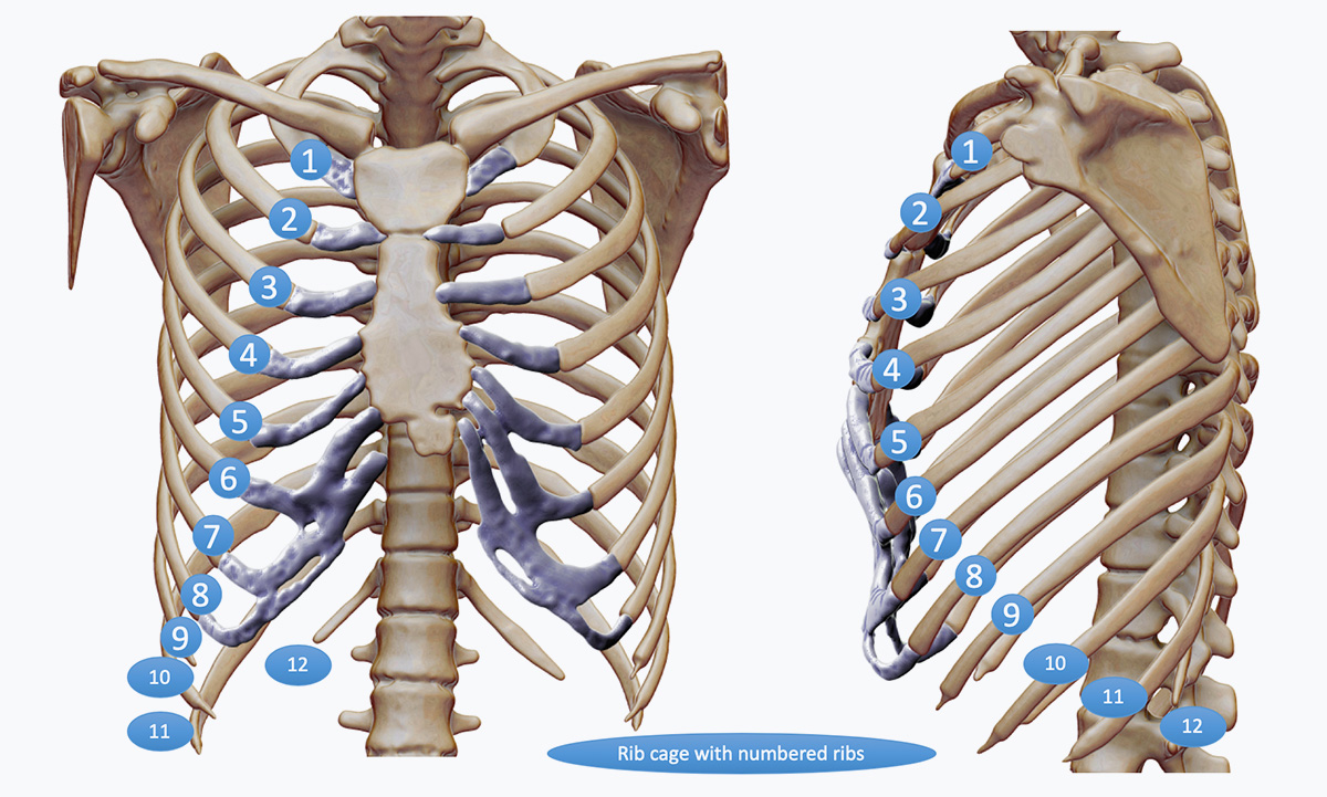
As the most common chest wall injury, it is usually caused by a direct blow or from a fall leading to bruising of the muscle of the chest wall and/or a rib break (fracture). If the rib or ribs are broken, what part of the rib depends on type of injury and which ribs are involved though typically it is the part of the rib called the neck (towards the back) or the shaft (side of the chest) of the rib that is broken. The ribs from top to bottom change shape and thickness. Generally, the ribs most vulnerable to injury are the 7th to 10th but any rib can be broken.
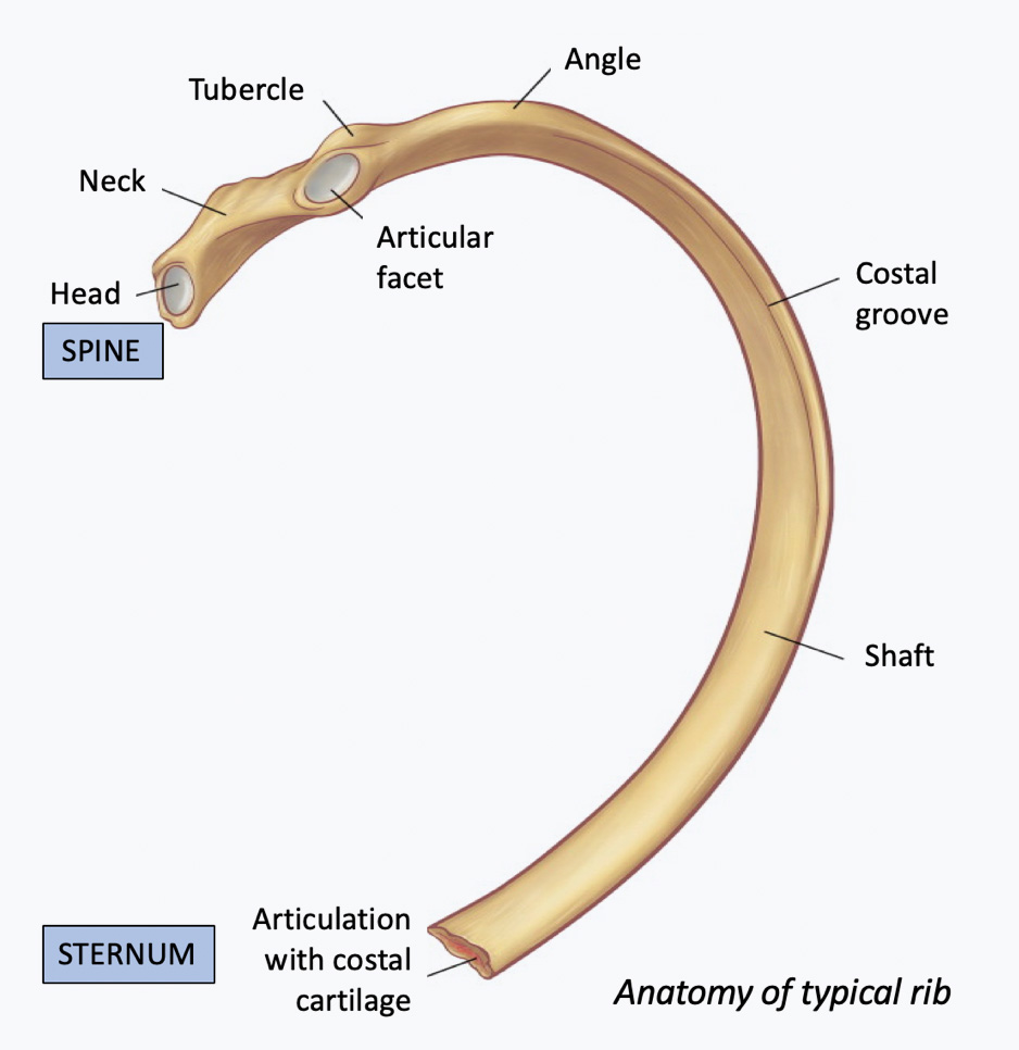
Anatomy of the typical rib
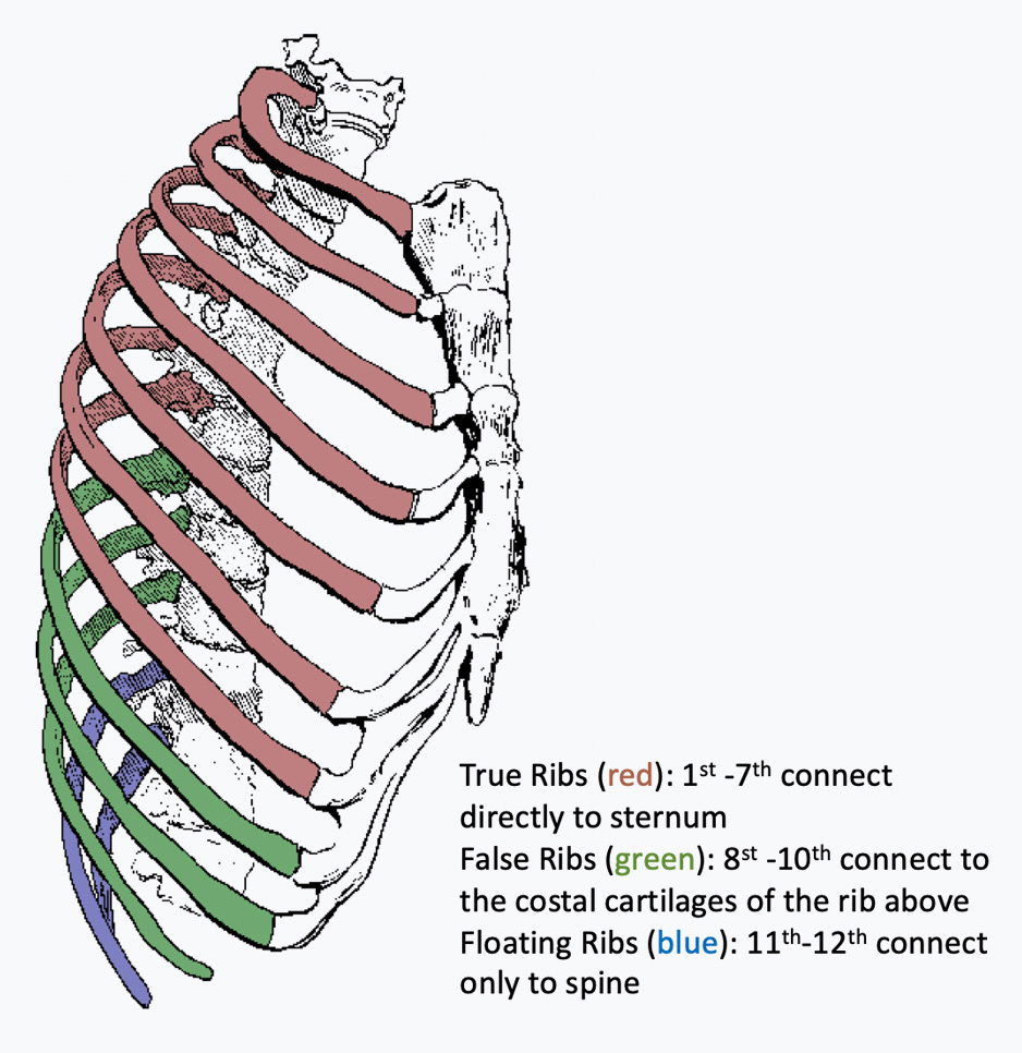
Rib cage showing the different types of rib depending on connection to sternum
Symptoms
Following the injury, depending on the severity of the injury it can cause immediate pain, swelling and bruising over the area injured and difficulty breathing. The chest typically hurts more when you take a deep breath in or with certain movements especially twisting. Coughing, sneezing or laughing also hurts more. If the rib is broken, you may feel or hear a ‘cracking’ sensation particularly on twisting. If the injury is minor, it can take up to 6 weeks for the pain and discomfort to settle. If the pain does not get better and is still affecting your breathing, there is a risk of developing a chest infection which can give you a persistent productive cough often with mucky green or yellowish sputum. Sometimes, particularly if there are more than one rib broken or the ribs were badly broken (displaced) the pain and discomfort can persist for months.
Diagnosis
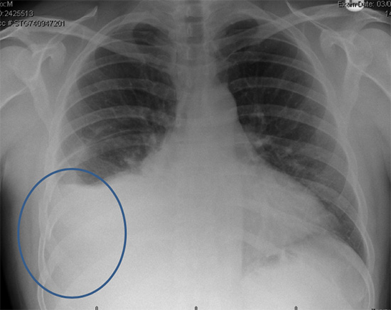
Chest x-ray showing several right-sided broken ribs and a collection of fluid or effusion in a middle-aged man with pain and breathlessness following a fall. He thought he might have ‘cracked’ a rib
The diagnosis of a rib injury is what doctors call a clinical one; that is taking a precise history of the injury coupled with a careful physical examination with a doctor familiar with chest wall injuries is usually all that is required, particularly if it’s a minor rib injury. There is no specific blood test unless an associated chest infection or other internal complication is suspected. Radiological assessment (chest x-ray) may be helpful to assess the severity of the rib injury and identify other associated problems such as fluid in the chest or a collapsed lung. If the injury is subtle occasionally a chest wall ultrasound may demonstrate a ‘hairline’ or partial rib fracture as well as identifying internal problems such a fluid (effusion), bruising of the lung (contusions) or lung collapse (pneumothorax).
The most sensitive radiological investigation particularly if more than one or two rib injuries is suspected, is a Chest CT scan. This allows the number and severity of the rib injuries to be clearly seen as well as identifying any other chest related injuries such as lung bruising or contusions.
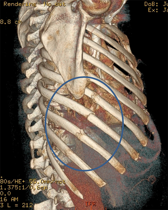
Following the Chest X-ray of the middle aged-man with pain and breathing issues he underwent a Chest CT scan and 3D reconstruction of his chest wall. This demonstrated 4 broken ribs from his fall.
Video of a Chest CT scan showing multiple right sided broken ribs and lung bruising (contusions) from a ski accident. Patient had a chest drain inserted.
Severity
Rib injuries vary significantly from a minor injury with associated pain and bruising to severe, multiple broken ribs and associated internal injuries. The type of injury and what happened is often the most useful guide to severity of rib injury.
Mild:
Type of injury: Minor such as knocking into object
Symptoms: Mild with discomfort and no obvious bruising or swelling though tender to touch associated with no breathing issues.
Diagnosis: Often clinical, requiring no investigations. If discomfort persists, consider Chest x-ray or if Chest x-ray normal and/or clinically suspicious (persistent pain and tenderness) consider Ultrasound assessment.
Moderate:
Type of injury: More serious such as a fall
Symptoms: Pain with breathing and obvious tenderness and/or bruising or swelling to the area of the chest.
Diagnosis: Clinical concerns of rib injury with or without signs of an obvious rib fracture/s, consider Chest x-ray or if associated with significant breathing concerns a Chest CT scan.
Severe:
Type of injury: Serious such as a fall from height (ladder or top of stairs) or road traffic accident as pedestrian, cyclist or motorcyclist, or driver
Symptoms: Serious constant Pain with tenderness, bruising and/or swelling to the area of the chest. Abnormal findings on chest examination.
Diagnosis: Clinically significant chest injury and usually requires Chest CT scan to assess chest wall injury and any associated internal injuries.
The classification of rib fractures is often based on the appearance of rib fractures on chest x-ray or commonly on Chest CT scan. They are described typically as:
Simple: Usually single sometimes described as a hairline fracture, the rib is not displaced (dislodged). May not be obvious on a chest x-ray. If localised pain persists, an ultrasound may detect fracture. However, even single or two rib fractures can be partially or completed displaced or fractured in more than one part of the rib.
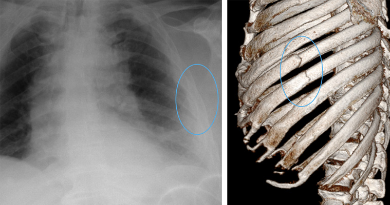
Chest X-ray (left) and CT reconstruction showing two simple non-displaced rib fractures (blue rings)
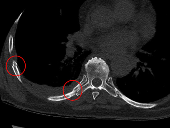
Chest CT demonstrating (in red circles) two non-displaced ‘simple’ rib fractures in different parts of the ribs
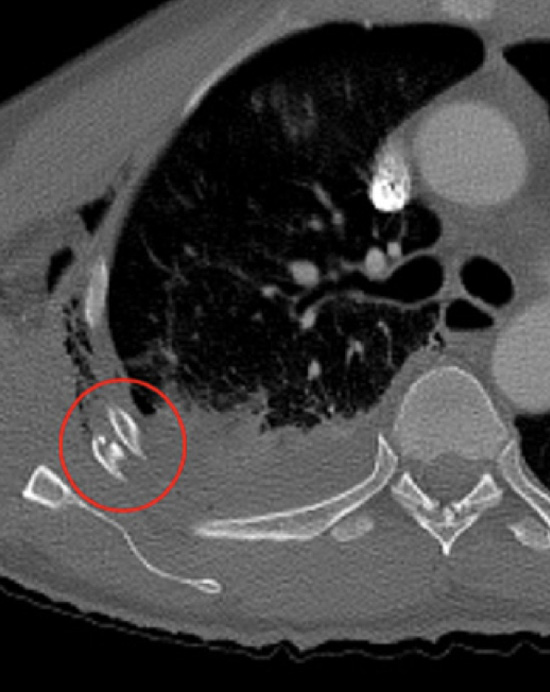
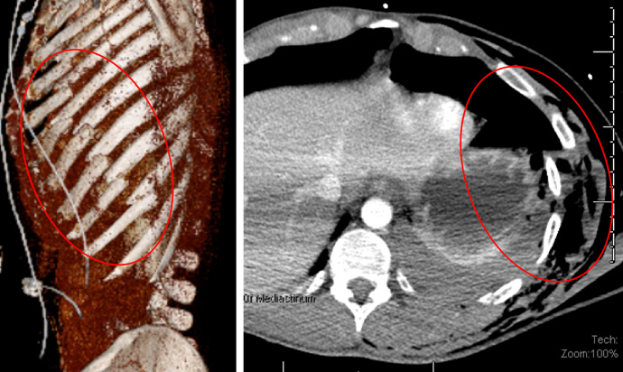
Complex: Typically, multiple rib fractures, usually displaced where the broken ends are misaligned. If multiple adjacent ribs are broken in multiple places, part of the chest wall can become separated, creating a segment which is free-floating and moves independently. This is called a ‘flail segment’ is serious and can affect breathing. This is often associated with lung bruising (contusions).
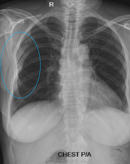
Chest X-ray showing multiple right sided rib fractures with several comminuted (broken in more than one place) fractures (blue ring)
CT chest reconstruction (left) showing multiple displaced complex rib fractures (right) and flail segment (red rings)
Chest CT demonstrating (red circle) a significantly displaced rib fracture with associated lung contusion
Complications of Rib Injury
Pain: Immediate (acute) and can be severe even after minor rib injuries, the area of rib injury is sore to touch and worse on certain movements. In most the pain will eventually settle, however occasionally it can persist and become chronic causing significant issues. Reasons of this include:
Failure to manage appropriately after the initial injury, no or not enough painkillers, appropriate rest, restrictions of activity and tailored return to normal activities.
Rib fractures or any bone that fails to heal properly can lead to conditions called mal-union, delayed healing or non-union. Symptoms of a fracture that is not healing normally include tenderness, swelling, and an aching pain that may be felt deep within the affected bone.
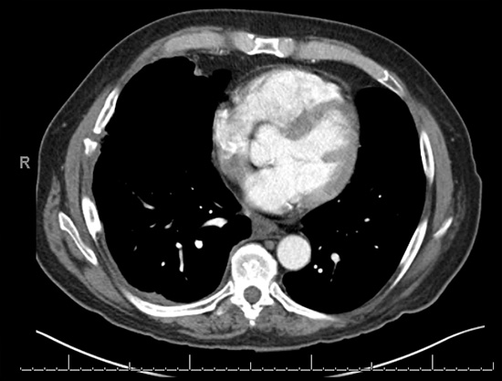
Chest CT scan showing a non-healed rib injury (non-union) and accompanying persistent pain
Rib injury can cause associated complex rib injury involving a junction between sternum and rib leading to dislocation or subluxation at the junction. For more information see complex chest wall injuries.
Breathlessness: Shortness of breath acutely is usually caused by the chest wall pain not allowing deep breaths to be taken, occasionally it can be associated with the lung collapsing after the injury; a build-up of fluid in the chest cavity (effusion) or even a developing chest infection (pneumonia). Chronically, on-going breathlessness can be due to chronic pain but also occasionally to complications of retained blood or fluid in the chest cavity which can trap the lung.
Internal injuries: Even relatively minor chest injuries can lead to internal injury to the lung (lung bruising (contusions), collapse (pneumothorax), effusions (blood or fluid) and rarely hernias (whether the lung or upper abdominal contents starts providing between broken ribs) or even a diaphragmatic (the muscle between the abdomen and the chest) hernia whereby the bowel contents slip into the chest from a hole or hernia in the diaphragm. Symptoms typically include on-going pain and breathlessness, swelling if a chest wall hernia and diagnosis requires a chest x-ray or even a chest CT scan.
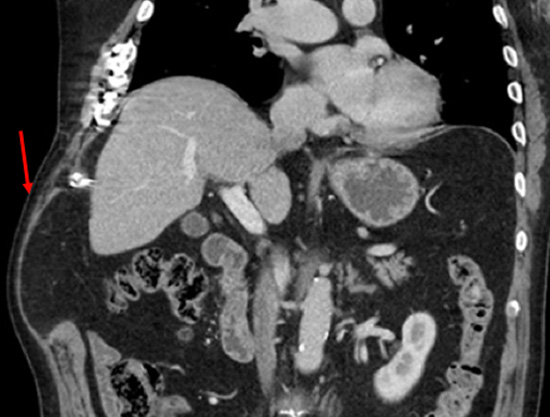
Chest CT scan showing a developing chest wall hernia (red arrow) following multiple rib injuries of the lower chest wall 2 years earlier
Treatment
Rib injuries can be treated either conservatively involving rest, restrictions of activities and painkillers, or through some form of intervention including targeted physical therapy, trigger point injections or a variety of surgical options. See Treatments.
